

How Oxidation Disrupts Cellular Communication
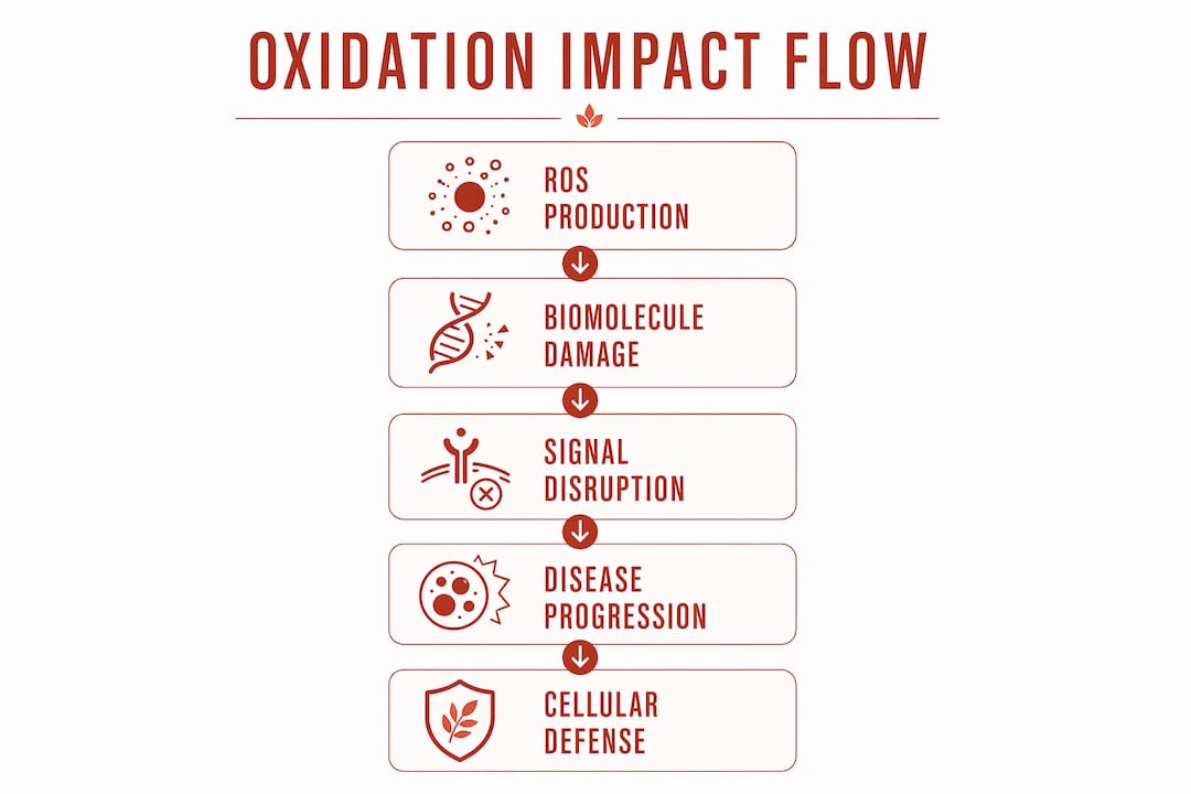
Oxidation disrupts cellular communication primarily by chemically modifying lipids, proteins, and DNA in ways that compromise the molecular machinery cells depend on to send and receive signals. When reactive oxygen species (ROS) accumulate beyond what the cell can manage, they trigger a cascade of irreversible molecular damage that impairs membrane integrity, disables receptor function, and corrupts the enzymatic switches that regulate signaling. The result is not just noise in the system. It is a breakdown of the coordinated biochemical conversation that keeps cells alive, differentiated, and responsive.
At the molecular level, the key disruptions look like this:
- Lipid peroxidation: ROS attack polyunsaturated fatty acids in cell membranes, generating reactive aldehydes like malondialdehyde (MDA) and 4-hydroxynonenal (4-HNE) that form adducts on proteins and DNA, distorting membrane fluidity and receptor geometry.
- Protein carbonylation: Oxidative modification of amino acid side chains, particularly cysteine and methionine residues, alters protein conformation and can permanently inactivate enzymes critical for signal transduction.
- DNA strand breaks and 8-oxoguanine (8-oxoG) formation: Oxidative lesions in DNA, including 8-oxoG, disrupt transcription and can produce truncated or mutated proteins that feed dysfunctional signals downstream.
- Phosphatase inactivation: ROS oxidize active-site cysteine residues in protein tyrosine phosphatases such as PTP1B and PTEN, removing the brakes on growth factor signaling and pushing cells toward uncontrolled proliferation.
- Receptor trafficking defects: Mitochondrial ROS impair exocyst membrane binding and endosomal recycling, blocking ligand delivery to the plasma membrane and silencing receptor-dependent pathways like Notch signaling.
- Regulated cell death activation: High, persistent ROS loads trigger apoptosis, ferroptosis, and innate immune cell activation, particularly in cardiovascular endothelium where redox imbalance is a primary driver of pathology.
Each of these mechanisms does not operate in isolation. They interact, amplify each other, and collectively erode the cell’s ability to communicate with precision.
How ROS function as essential signaling molecules under normal conditions
Before exploring what goes wrong, it helps to understand what ROS are supposed to do. The field has moved decisively away from the older model that treated all oxidation as damage. Today, researchers distinguish between oxidative eustress, the physiological use of ROS as second messengers, and oxidative distress, the excessive, uncontrolled oxidant challenge that breaks signaling and induces disease.

Hydrogen peroxide (H2O2) sits at the center of this distinction. It acts as a key second messenger by reversibly oxidizing cysteine thiolates on target proteins, a process precise enough to function like a molecular switch. Unlike superoxide, H2O2 is relatively stable, diffuses across membranes through specialized aquaporin channels called peroxiporins, and can be produced and degraded enzymatically, giving cells fine-tuned control over its concentration and duration.
Redox signaling operates through several well-characterized pathways:
- MAPK pathway: ROS activate MAP kinases by oxidizing upstream phosphatases, shifting the balance toward proliferative and stress-response gene expression.
- PI3K/AKT pathway: Oxidative inactivation of PTEN, a lipid phosphatase, amplifies PI3K signaling and promotes cell survival and growth.
- NF-κB pathway: ROS modulate IκB kinase activity and NF-κB nuclear translocation, coordinating inflammatory gene expression in immune cells.
- Nrf2/KEAP1 axis: Oxidation of KEAP1 cysteine residues releases Nrf2 to translocate to the nucleus and drive antioxidant gene expression, a feedback loop that protects cells from oxidative overload.
Spatial compartmentalization matters enormously here. ROS signals are not broadcast uniformly across the cell. Redoxosomes, specialized redox-active endosomes that form at the plasma membrane, concentrate H2O2 near specific receptor complexes, allowing localized oxidative modifications without global cellular damage. This microenvironment control is what separates a productive redox signal from a destructive one.
Cell-to-cell redox communication adds another layer. Gap junctions transfer redox-active molecules directly between connected cells via connexons. Extracellular vesicles (EVs), including exosomes, carry redox-active enzymes like peroxiredoxins (Prx1, Prx2) and protein disulfide isomerases to neighboring or distant cells, influencing their redox state without direct contact. The human proteome contains a large number of cysteine residues, with a proportion of them oxidized in any given cell type, underscoring how pervasive and structured redox regulation actually is.
Pro Tip: When designing redox signaling experiments, account for subcellular compartmentalization. A bulk measurement of cellular H2O2 will miss the localized microdomain signals that drive pathway-specific responses. Genetically encoded biosensors like roGFP-Grx reporters offer compartment-specific resolution that bulk assays cannot.
What molecular mechanisms drive oxidative damage to biomolecules?
Understanding the chemistry behind oxidative damage clarifies why it is so disruptive to cellular communication. ROS originate from several intracellular sources, each with distinct spatial and temporal profiles.
Primary ROS sources
- Mitochondrial electron transport chain (ETC): Complexes I and III are the main sites of superoxide (O2•−) generation. MnSOD in the mitochondrial matrix rapidly converts O2•− to H2O2, which then diffuses into the cytoplasm to participate in signaling or cause damage.
- NADPH oxidases (NOXs): NOX family enzymes deliberately produce O2•− at the plasma membrane and in endosomes. NOX2 in phagocytes generates the oxidative burst for pathogen killing; NOX4 produces H2O2 constitutively in vascular cells, contributing to both physiological signaling and, when overactivated, endothelial dysfunction.
- Endoplasmic reticulum (ER): Protein disulfide isomerase (PDI) and Ero1 generate H2O2 as a byproduct of oxidative protein folding, linking ER stress to cellular redox state.
- Monoamine oxidase (MAO): Located on the outer mitochondrial membrane, MAO generates H2O2 during neurotransmitter catabolism. Exacerbated MAO activity in pathological conditions contributes to cardiovascular and neurodegenerative disease.
- p66Shc: This oxidoreductase produces ROS by oxidizing cytochrome c using mitochondrial reducing equivalents, playing a documented role in carcinogenesis and aging.
Damage to lipids, proteins, and nucleic acids
| Macromolecule | Oxidative Modification | Key Products | Functional Consequence |
|---|---|---|---|
| Membrane lipids | Lipid peroxidation | MDA, 4-HNE | Membrane rigidity, receptor dysfunction, protein adducts |
| Proteins | Cysteine/methionine oxidation, carbonylation | Sulfenic acids, carbonyls | Enzyme inactivation, misfolding, aggregation |
| DNA/RNA | 8-oxoG formation, strand breaks | 8-oxoguanine, abasic sites | Transcriptional errors, truncated proteins |
| Signaling enzymes | Active-site cysteine oxidation | Sulfinic/sulfonic acids | Irreversible phosphatase inactivation |
Lipid peroxidation deserves particular attention because its products travel. MDA and 4-HNE are electrophilic aldehydes that diffuse from the membrane and form covalent adducts on distant proteins and DNA bases, extending oxidative damage well beyond the initial site of ROS production. Both are validated biomarkers for oxidative cellular damage in clinical and research settings.
Protein oxidation is similarly far-reaching. Cysteine residues are the primary redox sensors because their thiol groups are uniquely reactive toward H2O2 and other oxidants. Reversible oxidation to sulfenic acid (Cys-SOH) can be repaired by thioredoxin or glutaredoxin systems. Irreversible oxidation to sulfinic (Cys-SO2H) or sulfonic acid (Cys-SO3H) permanently disables the protein. Methionine oxidation to methionine sulfoxide is reversible by methionine sulfoxide reductases, but chronic oxidative stress overwhelms this repair capacity.
mRNA oxidation to 8-oxoG is a particularly underappreciated mechanism in neurodegeneration. Oxidized mRNA produces truncated or mutated proteins that accumulate in neurons, contributing directly to the pathology seen in Alzheimer’s and Parkinson’s diseases. 8-oxoG in mRNA serves as a biomarker for nucleic acid oxidative damage and is detectable in cerebrospinal fluid, making it a candidate for noninvasive disease monitoring.
How does oxidative stress connect to disease progression through signaling pathways?
The transition from localized redox signaling to systemic disease happens when oxidative modifications shift from reversible regulation to irreversible dysfunction. Three disease contexts illustrate this progression with particular clarity.
Cardiovascular disease
Endothelial cells are exquisitely sensitive to redox imbalance. Under physiological conditions, nitric oxide (NO) produced by endothelial nitric oxide synthase (eNOS) maintains vascular tone and suppresses inflammation. Excess ROS, particularly superoxide, rapidly reacts with NO to form peroxynitrite (ONOO−), depleting NO bioavailability and uncoupling eNOS. The result is endothelial dysfunction: impaired vasodilation, increased leukocyte adhesion, and a pro-inflammatory signaling environment that accelerates atherosclerosis. Oxidative inactivation of protein phosphatases in vascular smooth muscle cells simultaneously amplifies growth factor receptor signaling, promoting the pathological proliferation that narrows arterial walls. You can read more about how oxidative stress causes inflammation and what that means for vascular health.

Neurodegeneration
The brain is particularly vulnerable to oxidative disruption for two reasons: it consumes a disproportionate share of the body’s oxygen, and neurons have relatively limited antioxidant capacity compared to other cell types. In Alzheimer’s disease, ROS-driven lipid peroxidation generates 4-HNE adducts on tau protein and amyloid precursor protein, accelerating their aggregation. Oxidized mRNA produces dysfunctional proteins that neurons cannot clear efficiently, compounding proteotoxic stress. In Parkinson’s disease, dopaminergic neurons in the substantia nigra are especially exposed because dopamine metabolism via MAO generates H2O2 constitutively, creating a baseline oxidative load that mitochondrial dysfunction can push past the tipping point.
Cancer
ROS-mediated inactivation of phosphatases PTP1B, PTEN, and PTPN11 removes critical negative regulators of growth factor signaling. With these brakes disabled, receptor tyrosine kinase pathways run unchecked, driving the uncontrolled proliferation that characterizes tumor development. ROS are implicated in the initiation of multiple cancers, including melanoma, leukemia, and carcinomas of the lung, colon, and breast. Paradoxically, cancer cells also exploit elevated ROS to activate pro-survival NF-κB signaling and to remodel the extracellular matrix, facilitating invasion and metastasis.
Cellular senescence and the SASP
Chronic oxidative stress activates the DNA damage response (DDR), which triggers the p53-p21 axis and blocks cyclin-dependent kinase 2 (CDK2), arresting the cell cycle. When oxidative damage persists, p16 upregulation maintains this arrest permanently, and cells enter a senescent state. Senescent cells then secrete a cocktail of inflammatory cytokines, chemokines, and growth factors called the senescence-associated secretory phenotype (SASP). NF-κB, activated partly through ROS-driven DDR signaling, drives SASP components including IL-6 and IL-8. The SASP propagates secondary senescence in neighboring cells, turning a local oxidative insult into a tissue-wide communication failure.
Key disease mechanisms driven by oxidative signaling disruption:
- Peroxynitrite formation depletes NO and uncouples eNOS in vascular endothelium
- Phosphatase inactivation (PTP1B, PTEN) amplifies growth factor receptor signaling in tumors
- 4-HNE adducts on tau and amyloid precursor protein accelerate neurotoxic aggregation
- DDR activation by oxidized DNA triggers p53-p21 cell cycle arrest and SASP secretion
- NF-κB hyperactivation sustains chronic inflammation across cardiovascular, metabolic, and oncological contexts
- Extracellular matrix remodeling driven by ROS facilitates cancer cell invasion
What recent advances are reshaping our understanding of redox biology?
The past few years have produced several conceptual shifts that go well beyond the classical “ROS causes damage” framework. These advances matter for anyone designing experiments or interpreting clinical data on oxidative stress.
The ROS wave as a conserved communication mechanism. Originally identified in flowering plants, the ROS wave is an autopropagating cell-to-cell signaling pathway in which an initiating cell activates extracellular superoxide production via NADPH oxidase homologs. The resulting H2O2 is sensed by neighboring cells, triggering their own enhanced ROS production and propagating the signal across tissues within minutes. Research now shows this mechanism exists across unicellular algae, amoeba, ferns, mosses, and animals, suggesting it evolved before the divergence of unicellular and multicellular life. In mammalian systems, this wave-like H2O2 propagation coordinates stress responses across cell populations in ways that bulk redox measurements completely miss.
Non-cell-autonomous mitochondrial ROS. A striking recent finding shows that mitochondrial ROS production in one cell can disrupt receptor trafficking in adjacent cells without those cells producing excess ROS themselves. ROS from dysfunctional mitochondria impair exocyst membrane binding and block endosomal recycling, preventing ligands like the Notch ligand Delta from reaching the plasma membrane. Remarkably, overexpressing the trafficking GTPase RAB11 restores ligand delivery and rescues normal Notch signaling, identifying a potential therapeutic target for diseases driven by mitochondrial dysfunction.
Extracellular vesicle-mediated redox signaling. Exosomes and microvesicles carry redox-active cargo including peroxiredoxins, thioredoxins, and protein disulfide isomerases to distant target cells. Prx4, the only peroxiredoxin isoform with a signal peptide, follows the classical secretory pathway and is detectable in plasma. Extracellular Prxs function as damage-associated molecular patterns (DAMPs), binding TLR4 and activating NF-κB signaling in recipient cells. This means oxidative stress in one tissue can alter the immune and redox state of distant tissues through vesicle-mediated communication.
Noninvasive redox biomarker detection. Diagnostic technologies now analyze redox changes in extracellular fluids including plasma, cerebrospinal fluid, and synovial fluid without invasive procedures. Extracellular thiol switches, redox proteins, and low-molecular-weight thiols like glutathione (GSH) serve as systemic monitors of oxidative disruption. This shift toward noninvasive monitoring is enabling translational medicine applications that were impractical when redox assessment required tissue biopsies.
Therapeutic challenges with antioxidants. Broad-spectrum antioxidant supplementation has repeatedly underperformed in clinical trials, and the reason is now better understood. Generic antioxidants cannot be delivered to the precise subcellular sites and time windows where oxidative damage occurs. Their reaction rates with ROS are also slower than those of endogenous antioxidant enzymes, limiting their in vivo efficacy. Enhancing the NRF2 pathway and other endogenous defense networks offers a more targeted strategy, though off-target effects remain a challenge. For a broader perspective on how antioxidants support natural healing, the emerging consensus favors supporting the body’s own enzymatic defenses over flooding the system with exogenous scavengers.
Pro Tip: When evaluating redox biomarkers in extracellular fluids, distinguish between reduced and oxidized thiol forms. Total glutathione measurements miss the redox ratio that actually reflects cellular oxidative state. Methods like HPLC with electrochemical detection or mass spectrometry-based thiol profiling give you the oxidized-to-reduced ratio that matters clinically.
Current research tools for studying oxidative impacts on cell signaling:
- Genetically encoded H2O2 biosensors (HyPer, roGFP-Grx) for real-time compartment-specific imaging
- Proximity ligation assays to detect oxidized protein complexes in situ
- Redox proteomics using iodoacetamide-based alkylation to capture reversible cysteine modifications
- Electron paramagnetic resonance (EPR) spectroscopy for direct radical detection
- Mass spectrometry-based lipidomics for MDA and 4-HNE adduct profiling
- Extracellular vesicle isolation combined with redox proteomics for intercellular signaling studies
Which molecular targets does oxidation hit hardest in cellular communication?
Oxidative stress does not damage cells randomly. It preferentially attacks molecules whose function depends on precise chemical geometry, and nowhere is that more consequential than in the proteins and lipids that mediate cellular communication.
Receptor tyrosine kinases and their regulators
Growth factor receptors like EGFR and PDGFR contain cysteine residues in their extracellular domains and intracellular kinase domains. Oxidation of these residues can either activate or inhibit receptor function depending on location and extent. More consequentially, the phosphatases that terminate receptor signaling, particularly PTP1B, PTEN, and SHP-2 (PTPN11), carry catalytic cysteine residues with unusually low pKa values, making them exquisitely sensitive to H2O2 oxidation. Even modest increases in cellular H2O2 inactivate these phosphatases, prolonging and amplifying downstream kinase signals. This is the molecular basis for ROS-driven tumor promotion across multiple cancer types.
G protein-coupled receptors (GPCRs) and second messengers
GPCRs are vulnerable to oxidative modification at extracellular cysteine residues that form structural disulfide bonds. Aberrant oxidation disrupts receptor conformation and ligand binding affinity. Downstream, oxidative stress impairs cyclic AMP (cAMP) signaling by oxidizing adenylyl cyclase and phosphodiesterase enzymes. Calcium signaling is similarly disrupted: ROS oxidize ryanodine receptors and IP3 receptors on the ER membrane, causing aberrant calcium release that dysregulates calmodulin-dependent kinases and downstream gene expression.
Ion channels and membrane potential
Voltage-gated potassium and sodium channels contain cysteine and methionine residues in their gating domains. Oxidation shifts their activation thresholds, altering membrane excitability in neurons and cardiomyocytes. In cardiac cells, oxidation of the ryanodine receptor RyR2 causes spontaneous calcium release that triggers arrhythmias, a direct line from molecular oxidation to organ-level communication failure.
Transcription factors as redox sensors
NF-κB, AP-1, and p53 all contain cysteine residues whose oxidation state modulates DNA binding. NF-κB is activated by ROS-mediated IκB degradation but requires reduction of its DNA-binding cysteine (Cys62 in the p50 subunit) for full transcriptional activity. This creates a biphasic redox dependence: moderate ROS activates NF-κB nuclear translocation, while severe oxidation inhibits its DNA binding. The Nrf2/KEAP1 system works similarly, with KEAP1 cysteine oxidation releasing Nrf2 to drive cytoprotective gene expression.
Lipid second messengers
Phosphatidylinositol 4,5-bisphosphate (PIP2) is a membrane lipid that serves as a precursor for IP3 and diacylglycerol (DAG), two classical second messengers. ROS suppress PIP5K activity, reducing PIP2 levels at the plasma membrane. This directly impairs receptor-mediated signaling that depends on PIP2 hydrolysis and disrupts the membrane scaffolding that organizes signaling complexes. Research in both Drosophila and human cancer cell lines confirms that ROS-producing cells show significantly reduced PIP2 levels, connecting mitochondrial dysfunction to broad second-messenger dysregulation.
How do antioxidant defense systems protect cellular signaling?
The cell’s antioxidant network is not a passive mop-up crew. It is an active, hierarchically organized system that shapes the amplitude, duration, and specificity of redox signals while preventing oxidative damage from spiraling out of control.
Enzymatic defenses
Superoxide dismutase (SOD) acts as the first responder in the antioxidant relay, converting superoxide to H2O2 before it can react with NO or iron to generate more damaging species. Three isoforms cover distinct compartments: SOD1 (cytoplasm and mitochondrial intermembrane space), SOD2/MnSOD (mitochondrial matrix), and SOD3 (extracellular space). At Tryrevivify, we’ve built our patented formula around superoxide dismutase precisely because of this foundational role. Supporting SOD activity addresses oxidative disruption at its earliest point, before downstream damage to signaling molecules accumulates.
Catalase converts H2O2 to water and oxygen in peroxisomes, providing a high-capacity sink for bulk H2O2 that would otherwise overwhelm thiol-based redox sensors.
Thioredoxin (Trx) and glutaredoxin (Grx) systems reduce oxidized protein cysteines, restoring signaling proteins to their active conformations. Thioredoxin reductase (TrxR) regenerates reduced Trx using NADPH, making these systems dependent on metabolic status. Peroxiredoxins (Prx1-6), which use Trx as their electron donor, function as both peroxide scavengers and redox relay proteins that transfer oxidation to downstream targets, enabling signal transduction rather than just damage prevention.
Glutathione (GSH) is the most abundant intracellular antioxidant, present at millimolar concentrations. It scavenges ROS directly and serves as a cofactor for glutathione peroxidases (GPx), which reduce lipid hydroperoxides and H2O2. The GSH/GSSG ratio is a widely used index of cellular redox state. Astrocytes release GSH to supply neurons with cysteine for their own GSH synthesis, a form of transcellular antioxidant support that protects neurons from the oxidative stress of high metabolic activity.
The Nrf2 pathway as a master regulator
The nuclear factor erythroid 2-related factor 2 (Nrf2) pathway coordinates the transcriptional response to oxidative stress. Under basal conditions, KEAP1 targets Nrf2 for proteasomal degradation. When ROS oxidize specific KEAP1 cysteine residues, Nrf2 escapes degradation, translocates to the nucleus, and drives expression of over 200 cytoprotective genes including those encoding SOD, catalase, GPx, Trx, and GSH biosynthetic enzymes. This makes Nrf2 a systems-level regulator of cellular redox homeostasis, not just a single antioxidant pathway.
The limitation of exogenous antioxidant supplementation becomes clear in this context. Flooding cells with vitamin C or vitamin E addresses only a narrow slice of the oxidative challenge and cannot replicate the spatially targeted, enzymatically precise protection that endogenous systems provide. Enhancing Nrf2 activity or directly supporting SOD function, as Tryrevivify’s formula does through its patented superoxide dismutase and prebiotic fiber combination, works with the cell’s own architecture rather than around it. Prebiotic fiber supports the gut microbiome, which influences systemic inflammation and oxidative burden through the gut-immune axis, adding a complementary layer of protection. For a deeper look at how metabolic oxidative stress develops and what it means for cellular health, the connection between gut health and systemic redox balance is increasingly well-supported.
Which diseases show the clearest link to oxidation-induced communication failure?
Several conditions now have mechanistic evidence detailed enough to trace the path from specific oxidative modifications to disrupted cellular signaling to clinical pathology.
Atherosclerosis and cardiovascular disease
Oxidized low-density lipoprotein (oxLDL) activates lectin-like oxidized LDL receptor-1 (LOX-1) on endothelial cells, triggering NF-κB-dependent expression of adhesion molecules (VCAM-1, ICAM-1) and chemokines that recruit monocytes into the arterial wall. Simultaneously, peroxynitrite-mediated eNOS uncoupling reduces NO production, impairing vasodilation and creating a self-reinforcing cycle of endothelial dysfunction and inflammation. Oxidative inactivation of protein phosphatases in smooth muscle cells amplifies PDGF receptor signaling, driving the pathological proliferation that contributes to plaque formation. The oxidative stress and stroke risk connection follows directly from this endothelial communication breakdown.
Type 2 diabetes and insulin resistance
Excess ROS in insulin-sensitive tissues, particularly skeletal muscle and adipose tissue, oxidize and inactivate insulin receptor substrate (IRS) proteins and the PI3K/AKT pathway components that mediate glucose uptake. Oxidative inactivation of protein tyrosine phosphatases in pancreatic beta cells disrupts the insulin secretion response to glucose. The resulting communication failure between circulating glucose levels and cellular uptake machinery is a core feature of insulin resistance. Oxidative stress variations in diabetes span multiple tissue types and involve distinct ROS sources, from mitochondrial overload in hyperglycemic conditions to NOX-driven inflammation in adipose tissue.
Alzheimer’s disease
The amyloid cascade in Alzheimer’s disease intersects with redox biology at multiple points. Amyloid-beta (Aβ) peptides generate ROS through metal ion reduction, and the resulting lipid peroxidation products (4-HNE, acrolein) form adducts on synaptic proteins, impairing neurotransmitter receptor function and synaptic plasticity. Oxidized mRNA in neurons produces truncated forms of proteins involved in synaptic maintenance, compounding the communication deficit. Mitochondrial dysfunction in affected neurons generates non-cell-autonomous ROS that disrupt receptor trafficking in neighboring cells, spreading signaling failure through the neural network.
Parkinson’s disease
Dopaminergic neurons in the substantia nigra face a unique oxidative challenge because dopamine auto-oxidizes to generate quinones and H2O2, and MAO-mediated dopamine catabolism adds to this baseline H2O2 load. Alpha-synuclein, the protein that aggregates in Lewy bodies, is particularly susceptible to oxidative modification, and 4-HNE adducts on alpha-synuclein accelerate its aggregation and impair its normal function in synaptic vesicle trafficking. The resulting disruption of dopamine release and receptor signaling in the nigrostriatal pathway is the direct mechanistic link between oxidative stress and the motor symptoms of Parkinson’s disease.
Cancer
Beyond the phosphatase inactivation mechanisms described earlier, oxidative stress drives cancer progression through epigenetic reprogramming. ROS-mediated DNA methylation changes silence tumor suppressor genes, while oxidative modifications to histone-modifying enzymes alter chromatin accessibility in ways that favor oncogene expression. The SASP from oxidatively stressed senescent cells creates a pro-tumorigenic microenvironment by secreting growth factors and matrix metalloproteinases that support tumor angiogenesis and invasion. Immune cell communication is also disrupted: oxidative stress in the tumor microenvironment impairs T cell receptor signaling and promotes immunosuppressive regulatory T cell differentiation, helping tumors evade immune surveillance. Supporting immune function through reduced oxidative stress is therefore relevant not just to general wellness but to the immune-oncology interface.
What experimental methods do researchers use to study oxidative impacts on signaling?
Studying how oxidation affects cell signaling requires tools that can capture events happening in milliseconds, in specific subcellular compartments, and at the level of individual protein residues. The methodological toolkit has expanded considerably in recent years.
Live-cell imaging with genetically encoded biosensors
Genetically encoded redox biosensors have transformed the field by enabling real-time, compartment-specific measurement of H2O2 and glutathione redox potential. HyPer and its improved variants (HyPer7, HyPerRed) are fluorescent proteins fused to the H2O2-sensing domain of OxyR, providing ratiometric H2O2 detection with subcellular resolution. roGFP-Grx1 reports the glutathione redox potential (EGSH) in specific compartments including the cytoplasm, mitochondrial matrix, and ER lumen. These sensors allow researchers to watch redox signals propagate through living cells in response to growth factors, stress stimuli, or mitochondrial dysfunction, capturing the spatial dynamics that bulk assays miss entirely.
Redox proteomics
Identifying which specific cysteine residues are oxidized under a given condition requires mass spectrometry-based approaches. The iodoacetamide (IAM) alkylation strategy blocks free thiols before cell lysis, preserving the oxidation state of cysteines at the moment of sample collection. Subsequent reduction and labeling of previously oxidized cysteines with a mass-tagged reagent allows their identification and quantification by LC-MS/MS. OxiTMT and isoTOP-ABPP are two established platforms for quantitative redox proteomics that can profile hundreds of oxidized cysteines across the proteome simultaneously.
Electron paramagnetic resonance (EPR) spectroscopy
EPR directly detects unpaired electrons in free radicals, making it the gold standard for identifying specific ROS species. Spin-trapping agents like DMPO (5,5-dimethyl-1-pyrroline N-oxide) react with short-lived radicals to form stable adducts detectable by EPR. This approach is particularly valuable for distinguishing superoxide from hydroxyl radical and for measuring ROS production in isolated mitochondria or membrane fractions.
Proximity ligation and co-immunoprecipitation under redox-preserving conditions
Detecting oxidized protein complexes in situ requires proximity ligation assays (PLA) with antibodies specific to oxidized residues, or co-immunoprecipitation performed under conditions that preserve disulfide bonds (non-reducing SDS-PAGE). These approaches reveal which signaling complexes form or dissociate in response to oxidative stress, connecting molecular oxidation events to pathway-level outcomes.
Extracellular vesicle isolation and redox cargo analysis
Isolating EVs by differential ultracentrifugation or size-exclusion chromatography, followed by redox proteomics of EV cargo, allows researchers to characterize the redox signals cells send to their neighbors. Combining EV isolation with functional assays in recipient cells connects the molecular content of vesicles to their biological effects on target cell signaling.
Noninvasive biomarker analysis in body fluids
For translational and clinical research, extracellular redox monitoring in plasma, urine, cerebrospinal fluid, and synovial fluid provides systemic information without tissue biopsy. HPLC with electrochemical detection measures GSH/GSSG ratios and cysteine/cystine ratios in plasma. Immunoassays and ELISA-based platforms quantify MDA, 4-HNE protein adducts, 8-oxoG in urine, and extracellular peroxiredoxins. These noninvasive methods are increasingly used in clinical trials to monitor oxidative burden and assess the efficacy of antioxidant interventions in real time.
Key Takeaways
Oxidation disrupts cellular communication through irreversible molecular modifications that disable signaling enzymes, corrupt receptor function, and propagate damage non-cell-autonomously through vesicles and ROS waves.
| Point | Details |
|---|---|
| ROS are dual-role molecules | Physiological H2O2 acts as a second messenger via reversible cysteine oxidation; excess ROS cause irreversible damage to signaling machinery. |
| Phosphatase inactivation drives disease | ROS oxidize catalytic cysteines in PTP1B, PTEN, and PTPN11, removing growth factor signaling brakes and promoting cancer and metabolic dysfunction. |
| Non-cell-autonomous ROS spread damage | Mitochondrial ROS from one cell impair exocyst binding and receptor trafficking in adjacent cells, disrupting Notch and other signaling pathways. |
| Endogenous defenses outperform supplements | The NRF2 pathway and SOD-based enzymatic defenses provide spatially targeted protection that broad-spectrum antioxidant supplements cannot replicate. |
| Noninvasive redox monitoring is now feasible | Extracellular thiol switches and redox proteins in plasma and cerebrospinal fluid enable systemic tracking of oxidative disruption without tissue biopsy. |
Support your cellular defenses with Tryrevivify

Understanding how oxidation disrupts cellular communication is the first step. Acting on it is where Tryrevivify comes in. Our patented daily supplement combines superoxide dismutase, the enzyme that neutralizes superoxide at the source, with prebiotic fiber that supports the gut-immune axis and reduces systemic oxidative burden. Rather than flooding your system with generic antioxidants that can’t reach the right subcellular compartments, Tryrevivify works with your body’s own enzymatic architecture to maintain the redox balance that healthy cellular signaling depends on.
Explore Tryrevivify and see how targeted cellular support translates into real, measurable protection against oxidative disruption.